
The cell (from the Latin word cellula, meaning ‘small room’) is the basic structural and functional unit of life forms. A Plant cell is covered by a cell wall that provides a definite shape to the cell. Many different organelles are present inside the cell wall, Which we will discuss below.
Plant Cell Define
Plant cells are eukaryotic cells found in green plants, photosynthetic eukaryotes of the kingdom Plantae, meaning a membrane surrounds their nucleus. A variety of membrane-bound organelles within the plant cell perform specialized roles to maintain the normal functioning of cells.
Plant cell Overview
At the most fundamental level, animals, fungi, and protists comprise a single eukaryotic cell. In contrast, bacteria and archaea each have a single prokaryotic cell rather than several eukaryotic cells. The cell walls, chloroplasts, and central vacuoles of plant cells distinguish them from other organisms’ cells.
Chloroplasts are organelles that play an essential role in the functioning of plant cells. These are the structures in plants responsible for photosynthesis, which convert the energy from the sun into glucose. The cells release carbon dioxide and expel oxygen from their bodies during this process.
Animals and other organisms require oxygen and glucose to survive. Plants are called autotrophs because they can prepare their food and can not take food from other organisms like animals.
Plant cells are photoautotrophic because they use the sun’s light energy to make glucose. While Heterotrophic organisms are those that get food from both plants and animals.
A plant cell’s cell wall and central vacuole work together to provide the cell with rigidity. The plant cell stores water in the central vacuole, which expands the vacuole into the cell’s sides. The cell wall then exerts a force known as turgor pressure against adjacent cell walls.
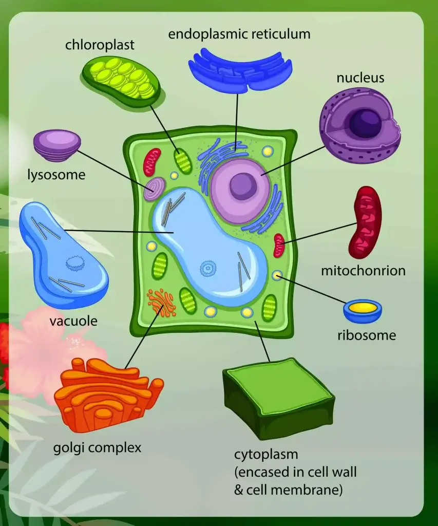
Plant Cell Diagram
Plant cells are quite different from animal cells because they perform different functions compared to each other. These differences can be understood when the cell is examined under an electron microscope.
Plant Cell Structure
The plant cell is distinct from the animal cell because of its rectangular shape and comparatively larger than the animal cell with a hard outer covering (cell wall). In contrast, the animal cell is round in shape and smaller than the plant cell.
Because of their eukaryotic nature, plant and animal cells share a few cell organelles, like mitochondria, ribosomes, nuclei, Endoplasmic Reticulum, etc. Plant cells have some structural organelles not seen in animals’ cells, like cell walls, vacuoles, plastids, Chloroplast, etc.
While animal cells also have some structures that are not seen in the plant cells, such as cilia and flagella, lysosomes, centrioles, etc.
Plant Cell consists of the following major cell organelles.
- Cell Wall
- Cytoskeleton
- Cell (Plasma) Membrane
- Plasmodesmata
- The cytoplasm
- Plastids
- Plant Vacuoles
- Mitochondria
- Endoplasmic reticulum (ER)
- Ribosomes
- Storage granules
- Golgi bodies
- Nucleus
- Peroxisomes
Plant Cell Wall
Discovery:
The cell wall was discovered by Robert Hooke in 1665.
Structure:
It is the outermost boundary of the plant cells. Each cell, whether isolated or occurring in tissues, has its cell wall. The chemical composition of the cell walls differs from specie too. E.g. plant cell wall is composed of cellulose, hemicellulose, and pectin.
The cell wall of fungi is composed of chitin, the Cell wall of bacteria is composed of murein, and the cell wall of algae is composed of glycoproteins and polysaccharides. Some other compounds, like i.e. lignin and cutin, are also found in the cell wall.
6000 Glucose monomers combine to form a cellulose chain. 100 cellulose chains combine to form Micelle. 20-40 micelles combine to form microfibrils, and 250 microfibrils combine to form microfibrils which are combined to form cell walls.
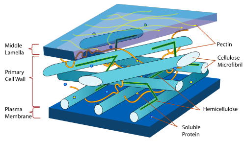
Cell walls have three fundamental layers, namely:
i) Middle lamella
ii) Primary wall
iii) Secondary wall.
i) Middle Lamella:
The middle lamella is a layer that cements the cell walls of two adjoining plant cells together. It is the first formed layer deposited at the time of cytokinesis. The cell plate formed during cell division develops into the middle lamella or labellum.
The middle lamella comprises calcium and magnesium pectate. In a mature plant cell, it is the outermost layer of the cell wall. It is usually 1 micrometer in thickness.
ii) Primary Wall:
This layer is formed between the middle lamella and plasma membrane in growing plant cells. It primarily comprises cellulose microfibrils within a gel-like matrix of hemicellulose fibers and pectin polysaccharides.
The primary cell wall provides the strength and flexibility needed for cell growth. It is usually 1-3 micrometers of thickness.
Secondary wall:
This layer is formed between the primary cell wall and plasma membrane in some plant cells. Once the primary cell wall has stopped dividing and growing, it may thicken to form a secondary cell wall. This rigid layer strengthens and supports the cell. In addition to cellulose and hemicellulose,
Some secondary cell walls contain lignin. Lignin strengthens the cell wall and aids in water conductivity in plant vascular tissue cells. It is usually 5-10 micrometers thick and consists of three layers: S1, S2, and S3.
FUNCTION OF CELL WALL
- It Provides a definite shape and structure to the cell.
- It Provides support and protection against infections.
- It helps in the transport of substances and communications in the cells.
- It prevents water loss.
- It helps in osmotic regulations.
- It prevents cells from rupturing due to turgor pressure.
- It provides mechanical protection from pathogens and insects.
- The cell wall stores carbohydrates for plant growth, especially in seeds.
- The cell wall sends signals for the cell to enter the cell cycle to divide and grow.
- It helps in the diffusion of gases in and out of the cell.
Also Read: Cell Wall
Cytoskeleton
The cytoskeleton is a structure that helps cells to keep their form and inner organization, and it additionally gives mechanical support that enables cells to carry out essential functions like division and movement. There is no single cytoskeletal component. it consists of the following components.
- Actin filaments, or microfilaments, are a network of parallelly arranged fibers. Actin filaments got their name because they are composed of tiny strands of actin proteins. They are the cytoskeleton’s thinnest filaments with a thickness of just 7 nanometers. They have a key role in the division of cell cytoplasm through cytokinesis, resulting in the formation of two daughter cells.. They also participate in cytoplasmic streaming, a process of cytosol flow all over the cell, transporting nutrients and cell organelles.
- Between actin filaments and microtubules, intermediate filaments with a diameter of 8–12nm, are found. Its role in plant cells is not entirely defined. The intermediate filaments’ role in the plant cells is not clearly understood. Still, it plays a role in maintaining the cell shape, structural support, and keeping tension.
- Microtubules are 23nm diameter hollow tubes formed of tubulins. Compared to the other two filaments, they are the largest filament. Unlike the role of the microtubule in cell division in the animal cell, the plant cell uses the microtubules to transport materials within the cell, and they are also used in forming the plant cell, cell wall.
Plant Cell Membrane
Cell Membrane was discovered by Swiss botanists Carl Naegeli and C. Crammer in 1855. The membrane is the second layer in plant cells below the cell wall, while it is the first in animal cells. Cell Membrane surrounds the cytoplasm and other organelles in it.
Structure:
In 1972, two scientists, S.J Singer and CL Nicolson, proposed a fluid mosaic model explaining the structure of the cell membrane.
Fluid Mosaic Model
According to this model, “Cell membrane comprises phospholipid by layer and proteins.
Some Cholesterol and carbohydrates are also present in the cell membrane. The phospholipid
bilayer forms a Fluid Sea in which proteins are floating.
Phospholipids:
Phospholipids have two ends.
1) Head:
Polar spherical heads are located over the cell surface. They have a phosphate group. They
are called hydrophilic (Water Loving)
2) Tail:
They are also called Nonpolar ends. They face each other in the middle of the bilayer. Tails of both
layers attract each other and repel water. So they are Hydrophobic. (Water Hating).
Cholesterol:
Cholesterol is also present in the cell membrane. They make the membrane less permeable
for water-soluble substances. It makes the membrane a rigid structure.
Carbohydrates:
Carbohydrates are present in two forms, i.e. Glycolipids and Glycoproteins.
Proteins:
The cell membrane contains two types of protein.
1) Peripheral Proteins:
These Proteins are also called Extrinsic Proteins. These are attached to the inner-outer membrane.
surface.
2) Integral Proteins:
These Proteins are also called Intrinsic Proteins. They are embedded in the lipid bilayer.. These
Proteins perform the following functions.
- Some link to sugar-Protein markers on the cell surface.
- Some move ions or molecules across the membrane.
- Some attach the membrane to the cell’s inner Cytoskeleton.
Functions of the Cell membrane
- It gives shape and protection to cells.
- Transport material into and out of the cell.
- Act as a receptor site, recognize chemicals, hormones, and neurotransmitters, and help signaling.
- The boundary separates the part of the cell from the outer environment.
- It helps in exocytosis and endocytosis.
- Regulates material moving into and out of the cell and from one part to another.
Also Read: Cell Membrane
Plasmodesmata
Plasmodesmata are small channels that connect the cytoplasm of adjacent plant cells. They allow the cells to communicate and exchange materials, such as hormones and nutrients, without losing their individual identities.
Plasmodesmata comprise a protein called desmoplakin, which forms a tube-like structure through which materials can pass.
The plant can regulate the channel’s diameter, allowing it to control the flow of materials between cells. Plasmodesmata are essential for the development and function of plant tissues and organs.
Cytoplasm:
The cytoplasm is a thick solution that fills each cell and is enclosed by the cell membrane. It is mainly composed of water, salts, and proteins. In eukaryotic cells, the cytoplasm includes all the material inside and outside the nucleus.
All organelles in eukaryotic cells, such as the nucleus, endoplasmic reticulum, and mitochondria, are located in the cytoplasm.
The portion of the cytoplasm that is not contained in the organelles is called the cytosol. Although cytoplasm may have no form or structure, it is highly organized. A framework of protein scaffolds called the cytoskeleton provides the cytoplasm and the cell with their structure.
Plastids
Plastids are a type of organelle found in plant and algae cells. They handle various functions, including the synthesis and storage of food, pigmentation, and the production of chemicals.
Plastids are like mitochondria in that they are both semi-autonomous organelles with their own DNA, but plastids are larger and have a greater variety of functions.
Examples of plastids include chloroplasts, which are responsible for photosynthesis; leucoplasts, which are involved in the synthesis and storage of starch, fatty acids, and other substances and chromoplast, which give different colors to fruits.

Plant Vacuoles
Vacuoles are another type of organelle found in plant cells. They are large, membrane-bound structures that serve several functions, including the storage of water, ions, and other molecules and regulating cell shape and turgor pressure.
Vacuoles also play a role in the digestion and degradation of macromolecules, and in plant cells, they are the site of pigment synthesis and storage. Vacuoles in plant cells are larger and more prominent than those in animal cells, and they can make up a significant portion of the cell’s volume.
Mitochondria
Mitochondria are organelles found in the cells of most eukaryotic organisms, including plants and animals. They are often referred to as the “powerhouses” of the cell because they generate most of its energy in the form of ATP.
Mitochondria have their own DNA and are semi-autonomous, meaning they can reproduce independently of the cell. They are typically rod-shaped and can be found in large numbers in cells that require a lot of energy, such as muscle cells.
Besides their role in energy production, mitochondria are also involved in other cellular processes, such as signaling, apoptosis, and the synthesis of lipids and certain amino acids.

Endoplasmic reticulum (ER)
The endoplasmic reticulum (ER) is an organelle found in eukaryotic cells. It is a network of flattened membrane-bound sacs and tubules involved in synthesizing, folding, and modifying proteins, lipids, and other molecules.
The ER has two main regions: the rough endoplasmic reticulum (RER), which is studded with ribosomes, and the smooth endoplasmic reticulum (SER), which lacks ribosomes.
The RER is involved in the synthesis and modification of proteins. In contrast, the SER is involved in the synthesis and modification of lipids, as well as the detoxification of drugs and other chemicals.
The ER is connected to the Golgi apparatus, which further processes and sorts the molecules that are produced by the ER.
Ribosomes
Ribosomes are small organelles found in the cells of all living organisms. They are the site of protein synthesis, where the information in mRNA molecules is used to produce proteins. Ribosomes consist of two subunits, large and small, which come together to form a complex.
The ribosomes read the information in the mRNA and use it to assemble the amino acids in a specific order to form a protein.
Storage granules
Storage granules are small organelles found in the cells of some Plants. They store various molecules, such as glycogen, lipids, and pigments. The molecules are stored in a concentrated form inside the granules, which helps to conserve space within the cell.
Storage granules can vary in size and shape depending on the organism and the molecules they store. For example, glycogen granules in liver and muscle cells are typically spherical, while lipid droplets in fat cells are often elongated.
Golgi bodies
Golgi bodies, also known as Golgi apparatus or Golgi complex, are membrane-bound organelles found in the cells of eukaryotic organisms. They are composed of stacks of flattened membrane sacs, which are involved in the sorting, modifying, and packaging of proteins and lipids as they are transported within the cell.
The Golgi complex receives newly synthesized proteins and lipids from the endoplasmic reticulum, modifies them, and then sorts them into vesicles for transport to their final destination.
The Golgi complex plays a critical role in the function of many cells, and defects in its structure or function can lead to various diseases.
Nucleus
The nucleus is a large organelle found in the cells of eukaryotic organisms. It is the cell’s control center, containing the genetic material (DNA) and directing the synthesis of proteins and other cellular activities.
A double membrane surrounds the nucleus called the nuclear envelope, which separates the contents of the nucleus from the rest of the cell.
The nucleus contains many structures, including the nucleolus, which is involved in the production of ribosomes, and chromatin, which consists of DNA and proteins and is the precursor to chromosomes during cell division.
The nucleus plays a central role in the function of the cell, and defects in its structure or function can have serious consequences for the organism.
Peroxisomes
Peroxisomes are small organelles found in the cells of eukaryotic organisms. They are similar to lysosomes, containing hydrolytic enzymes, but peroxisomes have different functions.
Peroxisomes contain enzymes that are used to break down fatty acids and other molecules through a process called oxidation.
This process produces hydrogen peroxide as a byproduct, which is then converted into water by another enzyme called catalase. Peroxisomes are essential for maintaining the health of the cell, and defects in their function can lead to various diseases.
Questions about Plant cell
Q. Why do plant cells need a cell wall and animal cells do not?
Ans: Plant cells need cell walls, whereas animal cells do not because plants need a rigid structure to grow up and out. All cells have cell membranes, and the membranes are flexible. So animal cells can have various shapes, but plant cells only have the shapes of their cell walls.
Q. What is a plant cell?
Ans: Plant cells are eukaryotic cells with a proper nucleus and specific organelles to carry out particular tasks. However, plant cells include several organelles unique from those found in other eukaryotic cells.
Q. Which organelles are found only in plant cells?
Ans: The chloroplast, cell wall, plastids, and a large central vacuole are the organelles limited to plant cells. The green pigment chlorophyll, which is found in the chloroplasts, is what causes photosynthesis to occur.
Q. What is the composition of a plant cell wall?
Ans: The cell wall of a plant is made up of cellulose. Cellulose is a long, linear polymer of several glucose molecules.
Where does photosynthesis occur in plant cells?
Inside the chloroplast of the plant cells, photosynthesis takes place. Chlorophyll, a green pigment, makes up chloroplast. The thylakoids of the chloroplast, which contain the pigment chlorophyll, are where the light reactions occur.
References
- 5% https://microbenotes.com/plant-cell
- 3% https://byjus.com/biology/plant-cell/
- 2% bing.com/videos
- 1% https://answers-all.com/popular/why-do-you-think-plant-cells-have-cell-walls
- 1% study.com/learn/lesson/site-of-protein-synthesis-in-cell.html
- 1% https://microbenotes.com/plastids-types-structure-and-functions
- 1% sciencing.com/vacuoles-definition-function-structure-137…