
The mitochondria (Gr.,mito=thread, chondrion =granle) are filamentous or granular cytoplasmic organelles of all aerobic cells of higher animals and plants and also of certain micro-organisms including Algae, Protozoa and Fungi.
These are absent in bacterial cells. They also contain specific DNA for cytoplasmic inheritance and ribosomes for protein synthesis.
History:
The mitochondria were first observed by Kolliker in 1850 as granular structures in the striated muscles. In 1888, he isolated them from insect muscles. Palade (1954) described the ultra-structure of cristae.
In 1963, Nass and Nass demonstrated the presence of DNA fibers in the matrix of mitochondria of embryonic cells. Attardi, Attardi and Aloni(1971) reported the 70S-type ribosomes inside the mitochondria.
Previously mitochondria have been known by various names such as fuchsinophilicgranules, paranasal bodies, plasmosomes, plastosomes, fila, vermicules, bioblasts and chondriosomes.
Distribution:
The mitochondria move autonomously in the cytoplasm, so they generally have a uniform distribution in the cytoplasm, but in many cells, their distribution is very restricted.
The distribution and number of mitochondria (and mitochondrial cristae) are often correlated with the type of function the cell performs.
Typically, mitochondria with many cristae are associated with mechanical and osmotic work situations where there are sustained demands for ATP and where space is at a premium, e.g., between muscle fibers, in the basal infolding of kidney tubule cells, and a portion of the inner segment of rod and cone cells of the retina.
Localization:
Often mitochondria occur in greater concentrations at work sites; for example, in the oocyte of Thyonebriaeus, rows of mitochondria are closely associated with RER membranes, where ATP is required for protein biosynthesis.
Mitochondria are particularly numerous in regions where ATP-driven osmotic work occurs, e.g., brush border of proximal kidney tubules, the infolding of the plasma membrane of dogfish salt glands and Malpighian tubules of insects, the contractile vacuoles of some protozoans (Paramecium).
Orientation:
The mitochondria have a definite orientation. For example, in cylindrical cells, the mitochondria usually remain orientated in a basal apical direction and lie parallel to the main axis. In leucocytes, the mitochondria remain arranged radially concerning the centrioles.
As they move about in the mitochondria, they form long moving filaments or chains. In contrast, in others, they remain fixed in one position where they provide ATP directly to a site of high ATP utilization, e.g., they are packed between adjacent myofibrils in a cardiac muscle cell or wrapped tightly around the flagellum of sperm.
Morphology:
Number:
The number of mitochondria in a cell depends on the type and functional state of the cell. It varies from cell to cell and from species to species. Certain cells contain exceptionally large numbers of mitochondria, e.g., the Amoeba Chaos Chaos contains 50,000; eggs of sea urchins contain 140,000 to 150,000; oocytes of amphibians contain 300,000. Certain cells, viz., liver cells of rats, contain only 500 to 1600 mitochondria.
Shape:
The mitochondria may be filamentous or granular in shape and may change from one form to another depending on the physiological conditions of the cells. Thus, they may be of club, racket, vesicular, ring or round shape.
Size:
Normally mitochondria vary in size from 0.5 µm to 2.0 µm and, therefore, are not distinctly visible under the light microscope. Sometimes their length may reach up to 7 µm.
Structure:
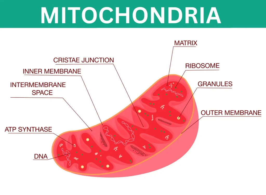
Each mitochondrion is bound by two highly specialized membranes that are important in its activities. Each of the mitochondrial membranes is 6 nm thick and fluid mosaic in ultrastructure.
The outer membrane is quite smooth and has many copies of a transport protein called porin, forming large aqueous channels through the lipid bilayer. Thus, this membrane resembles a sieve permeable to all molecules of 10,000 daltons or less, including small proteins.
Inside and separated from the outer membrane by a 6–8 nm wide space is present the inner membrane (Fig.10.4). The inner membrane is not smooth but is impermeable and highly convoluted, forming a series of infoldings, known as cristae, in the matrix space.
Mitochondrial Membrane Chambers:
Mitochondria are double membrane envelopes in which the inner membrane divides then mitochondrial space into two distinct chambers:
- The outer compartment is the peri-mitochondrial space or the inter-membrane space between the outer and inner membranes. This space is continuous into the core of the crests or cristae.
- The inner compartment, inner chamber or matrix space, is filled with a dense, homogeneous, gel-like proteinaceous material called mitochondrial matrix. The mitochondrial matrix contains lipids, proteins, circular DNA molecules, 55S ribosomes and specific granules related to mitochondria’s ability to accumulate ions.
Cristae:
In general, the cristae of plant mitochondria are tubular. In contrast, those of animal mitochondria are lamellar or plate-like (Hall, Flowers and Roberts, 1974). Still, in many Protozoa and steroid-synthesizing tissues, including the adrenal cortex and corpus luteum, they occur as regularly packed tubules (Tyler, 1973).
The cristae greatly increase the inner membrane area so that in liver cell mitochondria, the cristae membrane is 3–4 times greater than the outer membrane area.
Some mitochondria, particularly those from the heart, kidney and skeletal muscles, have more extensive cristae arrangements than liver mitochondria. In comparison to these, other mitochondria (e.g., from fibroblasts, nerve axons and most plant tissues) have relatively few cristae.
For example, mitochondria in epithelial cells of carotid bodies (or glomus carotica, which are chemoreceptors, sensitive to changes in blood chemistry and lie near the bifurcations of carotid arteries) have only four to five cristae and mitochondria from non-myelinated-axons-of rabbit brain have only a single crista.
Chemical Composition:
The gross chemical composition of the mitochondria varies in different animal and plant cells. However, the mitochondria contain 65 to 70 percent proteins, 25 to 30 percent lipids, 0.5 percent RNA and a small amount of DNA.
Lipids:
The lipid contents of the mitochondria are composed of 90 percent phospholipids (lecithin and cephalin), 5 percent or less cholesterol and 5 percent free fatty acids and triglycerides.
The inner membrane is rich in one type of phospholipid, called cardiolipin, which makes this membrane impermeable to a variety of ions and small molecules (e.g., Na+, K+, Cl—, NAD+, AMP, GTP, CoA and so on).
The outer mitochondrial membrane has a typical ratio of 50 percent proteins and 50 percent phospholipids of ‘unit membrane’. However, it contains more unsaturated fatty acids and less cholesterol.
It has been estimated that in the mitochondria of the liver, 67 percent of the total mitochondrial protein is located in the matrix, 21 percent is located in the inner membrane, 6 percent in the outer membrane, and 6 percent in the outer chamber.
Each of these four mitochondrial regions contains a special set of proteins that mediate distinct functions:
Enzymes of outer membrane:
Besides porin, other proteins of this membrane include enzymes involved in mitochondrial lipid synthesis and those enzymes that convert lipid substrates into forms metabolized in the matrix.
Certain important enzymes of this membrane are mono-amine oxidase, rotenone-insensitive NADH-cytochrome-C-reductase, kynurenine hydroxylase, and fatty acid CoA ligase.
Enzymes of intermembrane space:
This space contains several enzymes that use the ATP molecules passing out of the matrix to phosphorylate other nucleotides. The main enzymes of this part are adenylate kinase and nucleoside diphosphokinase.
Enzymes of inner membrane:
This membrane contains proteins with three types of functions:
- Those that carry out the oxidation reactions of the respiratory chain; 2. An enzyme complex called ATP synthetase makes ATP in the matrix.
- Specific transport proteins regulate the passage of metabolites into and out of the matrix.
- The important enzymes of the inner membrane are enzymes of electron transport pathways.
Enzymes of the mitochondrial matrix:
The mitochondrial matrix contains a highly concentrated mixture of hundreds of enzymes, including those required for the oxidation of pyruvate and fatty acids and the citric acid cycle or Krebs cycle.
The matrix also contains several identical copies of the mitochondrial DNA, special 55S mitochondrial ribosomes, tRNAs and various enzymes required to express mitochondrial genes.
Thus, the mitochondrial matrix contains the following enzymes: malate dehydrogenase, isocitrate dehydrogenase, fumarase, aconitase, citrate synthetase, α-keto acid dehydrogenase, and β-oxidation enzymes.
Moreover, the mitochondrial matrix contains different nucleotides, nucleotide coenzymes, and inorganic electrolytes K+, HPO4—, Mg++, Cl —, and SO4—.
Functions:
The mitochondria perform the most important functions, such as oxidation, dehydrogenation, oxidative phosphorylation and the respiratory chain of the cell. Their structure and enzymatic system are fully adapted for their different functions.
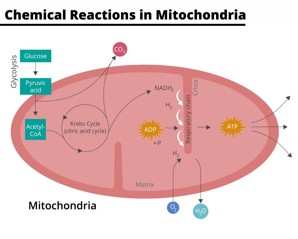
They are the actual respiratory organs of the cells where the foodstuffs, i.e., carbohydrates and fats, are completely oxidized into CO2 and H2O.
During the biological oxidation of carbohydrates and fats, a lot of energy is released, which the mitochondria utilize to synthesize the energy-rich compound known as adenosine triphosphate or ATP.
Because mitochondria synthesize energy-rich compound ATP, they are also known as “powerhouses” of the cell. In animal cells, mitochondria produce 95 percent of ATP molecules; the remaining 5 percent is produced during anaerobic respiration outside the mitochondria. In plant cells, ATP is also produced by the chloroplasts.
Adenosine triphosphate or ATP:
The ATP consists of a purine base adenine, a pentose sugar ribose and three molecules of the phosphoric acids. The adenine and ribose sugar collectively constitute the nucleoside adenosine which, by having one, two or three phosphate groups, forms the adenosine monophosphate (AMP), adenosine diphosphate (ADP) and adenosine triphosphate (ATP) respectively.
In ATP, the last phosphate group is linked with ADP by a special bond known as an “energy-rich bond” because when the last phosphate group of the ATP is released, a large amount of energy is released. Besides ATP, other energy-rich chemical compounds have recently been active in cellular metabolism.
These are cytosine triphosphate (CTP), uridine triphosphate (UTP) and guanosine triphosphate (GTP). However, these compounds derive energy from the ATP by nucleoside diphosphokinases.
The energy for producing ATP or other energy-rich molecules is produced during the breakdown of food molecules, including carbohydrates, fats and proteins (catabolic and exergonic activities).
Oxidation of carbohydrates:
The carbohydrates enter the cell as monosaccharides, such as glucose or glycogen. These hexose sugars are first broken down into a 3-carbon compound (pyruvic acid) by a series of chemical reactions known by many enzymes.
The pyruvic acid enters the mitochondria to oxidize into CO2 and water completely. The reactions which involve the oxidation of glucose into CO2 and water are known to form metabolic pathways, and they can be grouped under the following heads:
- Glycolysis or Embden-Meyerhof pathways (EMP) or Embden-Meyerhof-Parnas pathways (EMPP);
- Oxidative decarboxylation;
- Krebs cycle; citric acid cycle or tricarboxylic acid cycle;
- Respiratory chain and oxidative phosphorylation.
Other Functions of Mitochondria
Besides ATP production, mitochondria serve the following important functions in animals:
Heat production or thermogenesis:
Some mammals, especially young animals and hibernating species, have a specialized brown fat tissue. This tissue, typically located between the shoulder blades, is especially important in temperature regulation; it produces large quantities of body heat necessary for arousal from hibernation.
The color of brown fat comes from its high concentration of mitochondria, which are sparse in normal fat cells. The mitochondria appear to catalyze electron transport as usual but are much less efficient at producing ATP.
Hence, a higher-than-usual fraction of the oxidatively released energy is converted directly to heat (non-shivering thermogenesis).
Biosynthetic or anabolic activities:
Mitochondria also perform certain biosynthetic or anabolic functions. Mitochondria contain DNA and the machinery needed for protein synthesis. Therefore, they can make less than a dozen different proteins.
The proteins so far identified are subunits of the ATPase, portions of the reductase responsible for the transfer of electrons from Co Q to the iron of Cyt c, and three of the seven subunits in cytochrome oxidase.
Accumulation of Ca2+ and phosphate:
Large amounts of Ca2+ and phosphate (PO4¯ ) tend to accumulate in the osteoblasts’ mitochondria in tissues undergoing calcification. In the microcrystalline, electron-dense deposits may become visible.
Sometimes, the mitochondria assume a storage function, e.g., the mitochondria of the ovum store large amounts of yolk proteins and transform them into yolk platelets.
Key Points
- 💡 Mitochondria are rod-shaped organelles that act as the cell’s power generators.
- 💡 They convert oxygen and nutrients into ATP, the energy-rich compound.
- 💡 Mitochondria have specific DNA and ribosomes for cytoplasmic inheritance and protein synthesis.
- 💡 They were first observed in 1850 and have been known by various names.
- 💡 Mitochondria have a distribution and number correlated with the cell’s function.
- 💡 They can be found in regions where ATP-driven work occurs.
- 💡 Mitochondria have a definite orientation and can form long-moving filaments or remain fixed in one position.
- 💡 The number, shape, and size of mitochondria vary depending on the cell type and species.
- 💡 Each mitochondrion has two highly specialized outer and inner membranes.
- 💡 The inner membrane is convoluted and forms infoldings called cristae.
- 💡 Mitochondria contain different compartments, such as the peri-mitochondrial and matrix spaces.
- 💡 The cristae greatly increase the inner membrane area.
- 💡 Mitochondria have specific enzymes and proteins in different regions, mediating distinct functions.
- 💡 They play crucial roles in oxidation, dehydrogenation, oxidative phosphorylation, and the respiratory chain of the cell.
- 💡 Mitochondria are often referred to as “powerhouses” of the cell.
- 💡 They produce the majority of ATP molecules in animal and plant cells.
- 💡 Mitochondria are involved in the breakdown of carbohydrates, fats, and proteins to produce ATP.
- 💡 They also have additional functions like heat production and biosynthetic activities.
- 💡 Mitochondria can accumulate calcium ions (Ca2+) and phosphate (PO4¯) in certain tissues.
References:
- CELL BIOLOGY: P.S. VERMA, V.K. AGARWAL
- Cell Biology: Organelle Structure and Function By David E. Sadava – 1993
- Essential Cell Biology by Bruce Alberts, Dennis Bray, and Karen Hopkin – 2013
It’s enormous that you are getting ideas from this piece of writing as well as from our argument made at this time.
I have been surfing online more than three hours today, yet I never found any interesting article like yours. It抯 pretty worth enough for me. In my view, if all website owners and bloggers made good content as you did, the web will be much more useful than ever before.