
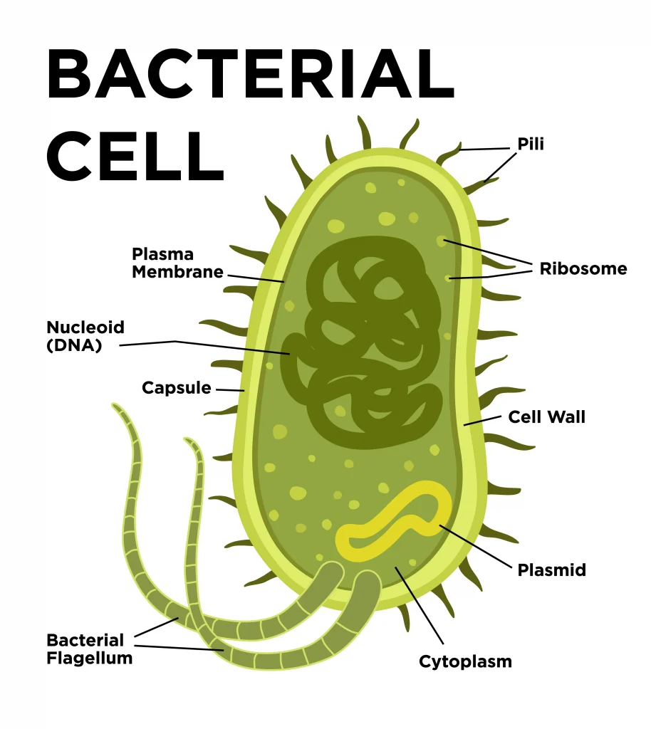
Bacteria are unicellular organisms with a simple structure. They are considered prokaryotes because of the absence of a well-formed nucleus. Structure and Function of a Typical Bacterial Cell with Diagram.
The cell structure includes a complex membrane, membrane-bound protoplast, cell walls, cytoplasm, and nucleoids that contain genetic material.
A bacterial cell’s outer layer or cell envelope comprises two components.
- A rigid cell wall.
- Underlying cytoplasmic or plasma membrane.
The cell envelope encloses the protoplasm, which comprises cytoplasm, cytoplasmic inclusions (mesosomes, ribosomes, inclusion granules, vacuoles) and a single circular chromosome of deoxyribonucleic acid (DNA). Besides these essential components, some bacteria may possess additional structures, such as capsules, flagella and fimbriae.
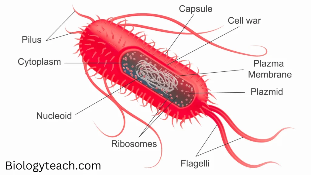
Structure and Function of a Typical Bacterial Cell
THE CELL WALL
The cell wall is a tough and rigid structure surrounding the bacterium like a shell. It weighs about 20-25% of the cell’s dry weight. The thickness of the Gram-negative cell wall is 10-25nm. The cell wall has the following functions:
- Accounts for the shape of the cell.
- Protects the cell against osmotic damage.
- Confer’s rigidity upon bacteria.
- It takes part in cell division.
- It possesses a target site for antibiotics, lysozymes, and bacteriophages. It carries bacterial antigens that are important in virulence and immunity.
| Gram-positive | Gram-negative | |
|---|---|---|
| Thickness | Thicker | Thinner |
| Periplasmic Space | Absent or small | Present |
| Lipids | Absent | Present |
| Teichoic Acid | Present | Absent |
| Peptidoglycan | 16-80 nm | 2 nm |
The rigid part of the cell wall is peptidoglycan, a mucopeptide (murein) composed of N-acetyl muramic acid and N-acetyl glucosamine molecules alternating in chains, cross-linked by peptide subunits. The cell walls of Gram-positive bacteria have a simpler chemical nature than Gram-negative bacteria.
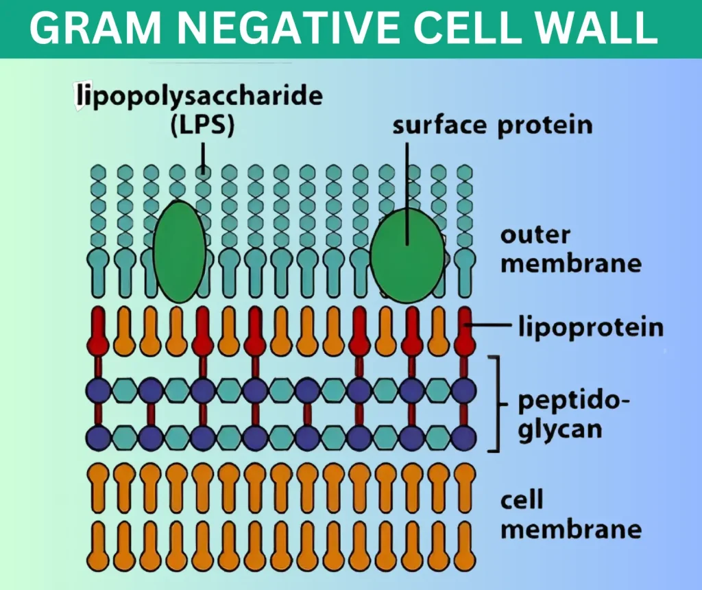
(i) Gram-negative Cell Wall
The Gram-negative cell wall is a complex structure with the following components.
- Lipoprotein layer. It connects the peptidoglycan to the outer membrane.
- Outer membrane: This contains certain outer membrane proteins (OMP) proteins. These are target sites for phages, antibiotics and bacteriocins.
- Lipopolysaccharide (LPS): This layer comprises lipid A attached to a polysaccharide. LPS makes up the endotoxin of gram-negative bacteria. The polysaccharide determines a major surface antigen, the O antigen. The toxicity (pyrogenicity, lethal effect, tissue necrosis) of bacteria is associated with lipid A.
- The periplasmic space is between the inner and outer membranes. It contains various binding proteins for specific substrates.
- Peptidoglycan.
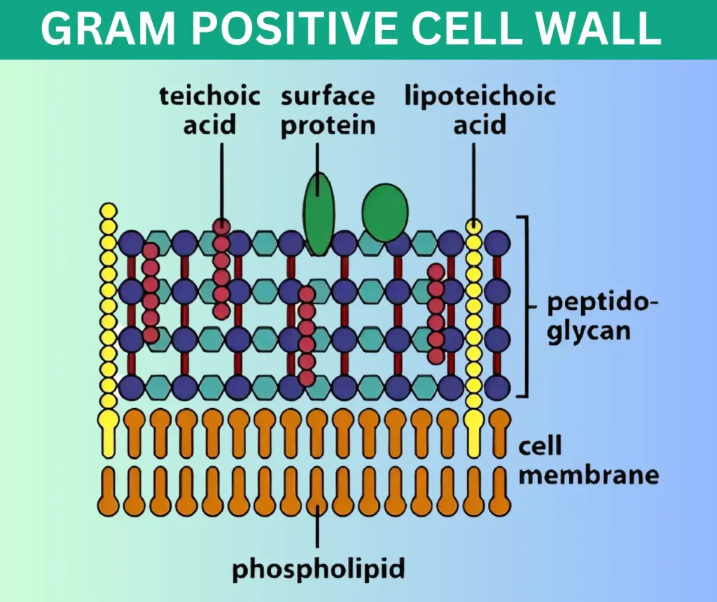
(ii) Gram-positive Cell Wall
- Peptidoglycan: This layer in Gram-positive bacteria is thicker (16-80nm) than in Gram-negative bacteria (2nm).
- Teichoic acid: Gram-positive cell wall contains a significant amount of teichoic acid, absent in Gram-negative bacteria. The teichoic acids make up the major surface antigens of Gram-positive bacteria. They are water-soluble polymers containing ribitol or glycerol polymers. Teichoic acids are of two types: cell teichoic acid and cell wall teichoic acid is covalently linked to peptGÄä7TTTe membrR¯iäéhoic acid to the cytoplasmic membrane.
- Other comFiEfi7. Certain Gram-positive cells also contain antigens such as proteins and
- Polysaccharides.
Cytoplasmic Membrane
It is a 5-10nm thick elastic semipermeable layer beneath the cell wall, separating it from the cytoplasm. Electron microscopy shows three layers constituting a unit membrane. Chemically, it comprises lipids and protein molecules. Sterols are absent, except in mycoplasma.
Cytoplasmic membrane act as an osmotic barrier. It is the site of many enzymes(permease, oxidase, polymerase) actively transporting selective nutrients.
It acts as a semipermeable membrane, controlling the inflow and outflow of metabolites to and from the protoplasm. In addition, it contains cytochrome oxidase, tricarboxylic acid cycle enzymes, and cell wall synthesis enzymes.
Cytoplasm
The bacterial cytoplasm is a colloidal system containing a variety of organic and inorganic solutes in a viscous watery solution. It lacks the mitochondria and the endoplasmic reticulum of eukaryotic cells. It contains ribosomes, mesosomes, vacuoles and inclusions.
The cytoplasm stains uniformly with basic dyes in young cultures.
(i) Ribosomes
These are the centers of protein synthesis. These are composed of ribosomal RNA (rRNA) and ribosomal proteins. Ribosomes are integrated into linear strands of mRNA to form polysomes, and it is at this site the code of mRNA is translated into peptide sequences.
They are 10-20nm in size with a sedimentation constant of 70 S (S for Svedberg units). Each 70 S unit consists of a 30 S and a 50 S subunit.
(ii) Intracytoplasmic inclusions
These are sources of stored energy and are present in some species of bacteria. Their function and significance are uncertain. They may be present as poly metaphosphate (volutin), lipid, polysaccharide (starch or glycogen) and sulfur granules.
They are most frequent in bacteria grown under nutritional deficiency conditions and disappear when deficient nutrients are supplied.
(iii) Mesosonws (chondroids)
Mesosomes are vesicular, multi-laminated or convoluted structures formed as invaginations of the plasma. They are the membrane into the cytoplasm. Principal centers of respiratory enzymes are analogous to the mitochondria of eukaryotes.
These are more prominent in Gram-positive bacteria. There are two types of mesosomes: septal and lateral.
The septal mesosome, an integral part of bacterial cells, plays a important role in the segregation of DNA and the formation of cross-walls during the process of binary fission. It remains attached to the bacterial chromosomes, facilitating these essential cellular processes.
Nucleus
The bacterial nucleus has no nuclear membrane or nucleolus. An ordinary microscope cannot show it but needs an electron microscope. The nuclear deoxyribonucleic acid (DNA) doesn’t contain any basic protein. The genetic material, known as genomic DNA, exists as a double-stranded circular structure.
It measures about I mm (1000µm) when straightened. The bacterial DNA is haploid, replicates by simple fission and maintains bacterial genetic characteristics. Some bacteria may possess extranuclear genetic material in the cytoplasm comprising DNA named plasmids or episomes.
The plasmid replicates autonomously. They are not essential for the life of the cell. Still, they may confer on the bacteria certain properties, such as drug resistance and toxigenicity, which make up a survival advantage to the bacteria.
Plasmids can be transferred between bacteria through conjugation or with the help of bacteriophages. In addition to these mechanisms, plasmids can also be passed on to daughter cells during cell division through binary fission.
Bacterial Capsul and Slime Layer
The amorphous viscid bacterial secretion surrounds some bacteria as their outermost layer. Where it disperses into the surrounding medium without clear boundaries, such as in Leuconostoc, it is referred to as a slime layer—a secretion that appears loose and un-demarcated.
When this secretion is organized into a sharply defined structure, as in Streptococcus pneumoniae (pneumococcus), it is known as the capsule. Capsules that are very thin and cannot be showed under the light microscope are called microcapsules, e.g., Nezsserza meningitidis.
The slime layer or capsule is-polysaccharide in nature, but it is a polypeptide in anthrax bacillus. Some bacteria may have a capsule and a slime layer (e.g., Streptococcus salivarius). Bacteria like Klebsiella secreting a large amount of slime produces mucoid growth on agar, with a stringy consistency when touched with the loop.
Functions
- Capsule enhances bacterial virulence by inhibiting phagocytosis. Loss of the capsule may render the bacterium avirulent. Bacteria tend to lose capsules on repeated subcultures.
- It acts as a protective covering against antibacterial substances such as bacteriophages and phagocyte enzymes.
- The capsular antigen is specific for bacteria and can be used to identify and type bacteria.
Capsulated Organisms
Streptococcus pneumoniae, Klebsiella sp., Bacillus anthracis, Haemophilus influenzae, Cryptococcus neoformans (a fungus).
Flagella
Flagella are cytoplasmic appendages protruding through cell wall. These are thread like structures composed of a protein (flagellin), 5-20 µm in length and 0.01-0.02µm in diameter. They are organ of locomotion. All motile bacteria, except spirochaetes, possess one or more
flagella.
Parts and Composition
Each flagellum consists of three parts.
(1) filament
(ii) hook
(iii) basal body
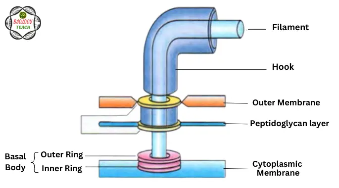
The filament Jies external to the cell and is connected to the hoök at the cell surface. The hook-basal body portion is embedded in the cell envelope. The basal body contains outer and inner rings by which the basal body is attached to the cytoplasmic membrane.
Outer rings are absent in Gram positive bacteria. The flagella is made up of protein (flaagellin) which is similar to myosin. Although chemical composition of different genera of bacteria is similar, they are antigenically different.
Specific flagellar antibodies are produced in high titres in response to antigenic stimulation of flagella. These antibodies are useful in serodiagnosis but are not protective.
Types According to Arrangement
- Monotrichous: Single polar flagellum (at one end) e.g. Vibrio cholerae.
- Amphitrichous: Single flagellum at both ends, e.g. Alcaligenes faecalis.
- Lophotrichous: Tuft of flagella at one or both ends, e.g. Spirilla.
- Peritrichous: Flagella arranged all around the cell, e.g. Salmonella typhi.
Fimbriae
These are hair-like. appendages projecting from the cell surface as shaight filaments. They are also called pilli. They are 0.1 to 1.0 µm in length and less than 10 nm thick (shorter and thinner than flagella). Fimbriae are found in some Gram negative bacteria.
Each bacterium possesses 100-500 fimbriae peritrichously. They are more numerous than flagella and are antigenic. In adition, they also best developed in freshly isolated strains an m 1quid cultures.
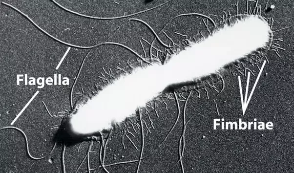
They disappear when subcultures are made on solid media. Fimbria is composed of protein called pilin. They are unrelated to motility and are found on motile and non-motile bacteria.
Types
There are three main types of fimbriae.
- Common pili: There are six types, depending on their morphology, number per cell, adhesive properties and antigenic nature.
- Sex or F (fertility) pili
- LiiiY Col I (colicin) pili
Functions
- Adhesion: Fimbriae are organs of adhesion. This property enhances the virulence of bacteria.
- Transfer of genetic material: Sex pili are present in male bacteria. These Pilli are longer (18-20 µm) and 1-4 in number. They help the male cells to attach with non-male (female) cells in forming “conjugation tubes” through which genetic material is believed to be transferred from the donor (male) to the recipient (female) cell.
Bacterial Spore
Spores are highly resistant resting stage formed in unfavourable environmental conditions presumed to be related to the depletion of exogenous nutrients. As bacterial spores are formed within the parent cell, these are called endospores.
Sporulation is not a method of reproduction.In the process of sporulation, each vegetative cell forms only one spore and during subsequent germination, each spore gives rise to only one vegetative bacterium.
Sporulation
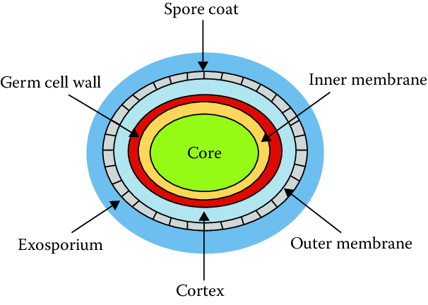
Bacterial cell undergoes spore formation in nutritionally deprived conditions and this process is called sporulation. Spore develops from a portion of protoplasm (forespore) near one end of the cell. The remaining part of cell is called sporangium.
Bacterial DNA replicates and divides into two DNA molecules. One of these is incorporated into forespore and other into sporangium. A transverse septum grows across the cell from the cell membrane. It divides forespore and sporangium.
The forespore is completely encircled by this septum as a double layered membrane. The inner layer becomes the spore membrane and the outer layer becomes thickened spore coat. Between the two layers is spore cortex.
Morphology of Spore
The clear area in the protoplasm of cell becomes gradually more opaque with condensation of nuclear chromatin forming the forespore. The cell membrane grows inward and forms spore wall around the core (forespore).
The inner-most layer of the spore wall forms the spore membrane from which the cell wall of future vegetative bacterium develops. Outside this membrane is thick layer, the cortex and a multilayered tough spore coat. Some spores _have an additional apparently rather loose, outercovering called exosporium.
Shape and Position of Spores
The precise position, shape and relative size of spore are constant within a particular species. Spores may be central, subterminal or terminal. They may be oval or spherical in shape.
The diameter of spore may be same or less than the width of bacteria (Bacillus), or may be wider than the bacillary body producing a distension or bulge in the cell ( Clostridiufn ).
Resistance
Bacterial spores are extremely resistant to ordinary boiling, disinfectants and heating. The high resistance of spores is due to high content of calcium and dipicolinic acid; low water content; The thick impervious cortex and spore coats; their low metabolic and enzymatic activity.
However, spores of all medically important bacteria are destroyed by autoclaving at 121 dgree celcious for 15 minute.
Germination
The process of conversion of a spore into vegetative cell under suitable conditions is known as germination. The spore loses its refractivity, and swells when transferred to conditions conducive to vegetative growth.
The spore wall is shed and the germ cell appears by rupturing the spore coat. The formation of vegetative bacterium occurs by elongation of the germ cell.
Key Points
- 🧫 Bacteria are unicellular organisms with a simple structure.
- 🏢 A typical bacterial cell resembles a plant cell and has a complex membrane, cell walls, cytoplasm, and nucleoids.
- 🧱 The cell envelope comprises a rigid cell wall and an underlying cytoplasmic or plasma membrane.
- 💪 The cell wall accounts for the cell’s shape, protects it against osmotic damage, confers rigidity, and plays a role in cell division.
- 🧪 Gram-positive and Gram-negative bacteria have differences in cell wall thickness, periplasmic space, lipids, teichoic acid, and peptidoglycan composition.
- 💦 The cytoplasmic membrane is a semipermeable layer beneath the cell wall, separating it from the cytoplasm, and is involved in transport and metabolic processes.
- 🔬 The cytoplasm contains ribosomes for protein synthesis, mesosomes involved in respiratory enzymes, and various inclusions such as stored energy sources.
- 🧬 The bacterial nucleus lacks a nuclear membrane and nucleolus, contains DNA in a circular form, and may have extranuclear genetic material called plasmids.
- 🎩 Bacterial capsules and slime layers are the outermost layers that enhance virulence and protect against antibacterial substances.
- 🚩 Flagella are protein-based appendages used for locomotion, and fimbriae (pili) are shorter hair-like appendages involved in adhesion and genetic transfer.
- 🌰 Bacterial spores are highly resistant resting stages formed in unfavorable conditions, and sporulation is the process of spore formation from vegetative cells.
FAQs
1. Can bacterial cells have more than one flagellum?
Yes, some bacterial cells can have multiple flagella, allowing them to exhibit more complex forms of movement.
2. Are all bacterial cell walls the same?
No, bacterial cell walls can vary in composition. Some bacteria have cell walls with a thick peptidoglycan layer, while others may have additional layers or different materials in their cell walls.
3. How do bacterial cells obtain nutrients from their environment?
Bacterial cells have various mechanisms to obtain nutrients. These include active transport, facilitated diffusion, and secretion of enzymes to break down complex molecules.
4. Can bacterial cells communicate with each other?
Yes, bacterial cells can communicate through a process called quorum sensing, where they release and detect chemical signals to coordinate group behaviors.
5. Are all bacterial cells harmful?
No, while some bacterial cells can cause diseases, many bacterial species are beneficial and play vital roles in natural ecosystems, nutrient cycling, and human health.
What is gram-negative cell wall?
Gram-negative bacteria have a thin peptidoglycan cell wall surrounded by an outer membrane that contains lipopolysaccharide. On the other hand, Gram-positive bacteria lack an outer membrane but have multiple layers of peptidoglycan that are significantly thicker than those found in Gram-negative bacteria.
References and Sources:
- Textbook Of Microbiology By C. P. Baveja
- Prokaryotic Cell Wall Compounds by Ajit Varma, Harald Claus, Helmut König
- https://www.ncbi.nlm.nih.gov/pmc/articles/PMC2857177/
- https://en.wikipedia.org/wiki/Gram-negative_bacteria
- https://byjus.com/question-answer/what-is-gram-negative-cell-wall/
- https://open.oregonstate.education/generalmicrobiology/chapter/bacteria-cell-walls/
- https://www.researchgate.net/figure/Structure-of-bacterial-spore_fig1_341354043