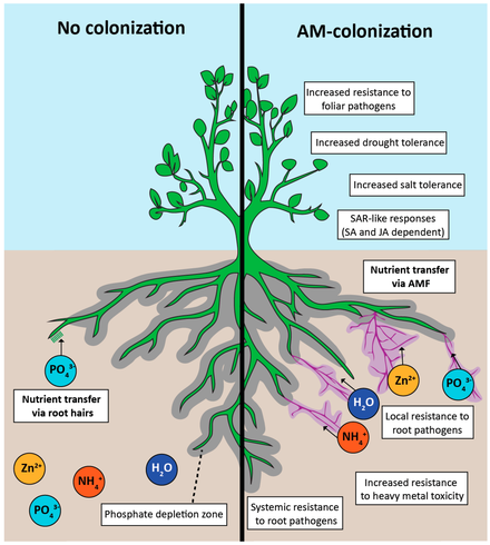
Uncover the hidden wonders of Arbuscular Mycorrhiza! From boosting plant growth to enhancing nutrient uptake, find out why this fungus is a game-changer for your garden.
Arbuscular mycorrhizas (AM) are the most common mycorrhizal type. They are formed in an enormously wide variety of host plants by obligately symbiotic fungi which have recently been reclassifi ed on the basis of DNA sequences into a separate fungal phylum, the Glomeromycota (Schüßler et al., 2001).
The plants include angiosperms, gymnosperms and the sporophytes of pteridophytes, all of which have roots, as well as the gametophytes of some hepatics and pteridophytes.
The name ‘arbuscular’ is derived from characteristic structures, the arbuscules which occur within the cortical cells of many plant roots and also some mycothalli colonized by AM fungi.

Mycorrhizal associations produced by Glomeromycotan fungi are known as arbuscular mycorrhizas, or vesicular-arbuscular mycorrhizas (formerly also endomycorrhizas, or endotrophic mycorrhizas) and are abbreviated as VAM here.An arbuscular mycorrhiza has three important components: the root itself, the fungal structures within and between the cells of the root and an extraradical mycelium in the soil.
Structures in Soil
Hyphae
A network of hyphae forms in the soil with thicker hyphae which function as conduits.
Absorptive hyphae
Thin highly branched hyphae are thought to absorb nutrients.
Spores
Large (for a fungus) asexual spherical structures (20-1000+ µm diameter) formed on hyphae in soil, or in roots.
Structures in Roots
Hyphae
These are non-septate when young and ramify within the cortex.
Arbuscules
Intricately branched haustoria in cortex cells.
Vesicles
Storage structures formed by many fungi.
Plant species involved:
It has been estimated that over 80% of all vascular plants form arbuscular mycorrhizas.
Arbuscular mycorrhizas have been identified in a broad spectrum of plants, including some non-vascular plants, ferns and other seedless vascular plants, groups within the gymnosperms including conifers (e.g., Thuja, Sequoia, Metasequoia), Ginkgo biloba, the cycads, and the majority of angiosperm families.
Brassicaceae (this family includes canola, mustards, cabbages, etc.) and the Chenopodiaceae (this family includes garden and sugar beets, spinach and the large genus Chenopodium), although even here arbuscular mycorrhizal associations have been reported for a few species.
Fungal species involved
The fungi involved are ubiquitous soil-borne organisms (Phylum Glomeromycota; Class Glomeromycetes) belonging to four orders: Archaeosporales, Paraglomerales, Diversisporales and Glomerales (Schüßler et al. 2001).
These genera, Glomus, Paraglomus, Sclerocystis, Scutellospora, Gigaspora, Acaulospora, Archaeospora, and Entrophospora, include approximately 150 species that are involved in the association.
All arbuscular mycorrhizal fungal species are obligate biotrophs, depending entirely on host plants for carbon compounds. This means that, unlike many ectomycorrhizal fungi, these fungi cannot be cultured in the absence of plants.
As a result, researchers have used root organ cultures and genetically transformed root cultures to maintain arbuscular mycorrhizal fungi and for use in experimental studies.
Structures and Developmental Stages:
The formation of arbuscular mycorrhizas involves a series of steps from the recognition of the root surface by the fungus to the formation of an appressorium, epidermal cell penetration, intraradical hyphal and arbuscular development, and, in some cases, the formation of vesicles.
Soil Hyphae:
Soil hyphae, also known as extraradical or external hyphae, are filamentous fungal structures which ramify through the soil. They are responsible for nutrient acquisition, propagation of the association, spore formation, etc.
VAM fungi produce different types of soil hyphae including thick “runner” or “distributive” hyphae as well as thin “absorptive” hyphae (Friese & Allen 1991). The finer hyphae can produce “branched absorptive structures” (BAS) where fine hyphae proliferate (Bago et al. 1998).
Hyphae of Scutellospora and Gigaspora species produce clustered swellings with spines or knobs called auxiliary cells.
Root Contact and Penetration
In order for the development of all internal structures to occur, fungal hyphae must contact either the surface of root epidermal cells (Figures) or root hairs (Figure), form specialized attachment structures called appressoria (swellings) and then penetrate the epidermal layer before reaching the cortex.
Hyphal Proliferation in the Cortex:
Aseptate hyphae spread along the cortex in both directions from the entry point to form a colony. The regions along the root at which appressoria form and where hyphae enter the epidermis are referred to as entry points.
Hyphae within roots are initially without cross walls, but these may occur in older roots. Gallaud (1905) observed that VAM associations in different species formed two distinctive morphology types, which he named the Arum and Paris series. Both types of associations are important in ecosystems (Smith & Smith 1997).
Coiling (Paris) Arbuscular Mycorrhizas:
In the Paris-type mycorrhiza, the intraradical hyphae pass from cell to cell, forming complex coils in both epidermal and cortical cells. Species of plants developing Paris-type mycorrhizas lack conspicuous intercellular spaces in the root cortex so that the growth of hyphae in the longitudinal direction of the root is slower than in the Arum-type.
Linear (Arum) Arbuscular Mycorrhizas:
In the Arum-type arbuscular mycorrhiza, the hypha that penetrates into the epidermis generally forms a coil either in the epidermal cell or first cortical cell layer (Figures) before it enters the intercellular spaces of the cortex .
In this type, roots can become rapidly colonized along the root axis due to the free movement of hyphae in the intercellular spaces.
Functions
Intraradical hyphae are able to initiate other fungal structures within the root. For example, in the Arum-type mycorrhiza, hyphae penetrate through the walls of cortical cells, branch repeatedly, and form arbuscules.
In the Paris-type, small branches develop laterally from the hyphal coils to form arbuscules (referred to as arbuscular coils. Intraradical hyphae may also initiate intracellular or intercellular vesicles.
Intraradical hyphae that persist in decaying root pieces in the soil are important sources of inoculum for the subsequent colonization of new roots.
Arbuscules
Arbuscules are intricately branched haustoria that formed within a root cortex cell. They were named by Gallaud (1905) because they look like little trees.
Development and Structure
The development of intracellular hyphae and arbuscules leads to dramatic changes in the organization of the cytoplasm in host cells. Cortical cells that were vacuolated prior to fungal penetration have an increased number of organelles such as mitochondria.
One of the most striking changes, however, is in the cytoskeleton Arbuscules start to form approximately 2 days after root penetration (Brundrett et al. 1985). They grow inside individual cells of the root cortex, but remain outside their cytoplasm, due to the invagination of the plasma membrane. Arbuscules are considered the major site of exchange between the fungus and host.
Functions
The highly branched nature of arbuscules and the presence of a periarbuscular membrane enveloping all of the fine branches provide a large surface area. Arbuscules are short-loved and begin to collapse after a few days, but hyphae and vesicles can remain in roots for months or years.
Intraradical vesicles:
Development and Structure
Vesicles are hyphal swellings in the root cortex that contain lipids and cytoplasm.Vesicles can develop from the tips of hyphae or from lateral branches, either within cells or in intercellular spaces of the root.
When vesicles form within root cells, they frequently enlarge to occupy the entire volume of the cell; they may assume the shape of the cell and even stretch the walls of the cells in which they occur
Vesicle formation usually follows that of arbuscules, indicating that the fungus may require arbuscular-derived sugars from the host prior to their formation. Vesicle development involves the swelling of a hyphal tip or lateral branch followed by modifications to the cytoplasm and cell wall.
The most characteristic cytological features of mature vesicles are the presence of lipid bodies and many nuclei. Bacteria also occur in vesicles of some species.
Functions
The storage of large amounts of lipid (as much as 58% of the dry mass of vesicles) indicates that vesicles are important storage structures and that they may act as propagules for the fungus.
Extraradical vesicles (auxiliary vesicles)
Development and Structure
Instead of intraradical vesicles, two genera, Gigaspora and Scutellospora, form auxiliary vesicles in the extraradical mycelium. These are initiated as lateral branches that rapidly expand into globose structures of varying colour and with ornamented walls.
Auxiliary vesicles can occur singly or in clusters. Wall ornamentation patterns are used as taxonomic characters. Each auxiliary vesicle stores large amounts of lipid, has vacuolar inclusions and is multi-nucleate. Bacteria may be present within the cytoplasm.
Functions
Very little is known about the role that auxiliary vesicles play in the life cycle of the fungus, although new hyphae have been observed growing from the subtending hyphae of these structures in Gigaspora margarita, suggesting that they may function in propagation.
Extraradical mycelium
Development and Structure
The extraradical mycelium of arbuscular mycorrhizas, like that of other mycorrhizas, has been difficult to study in situ because of the heterogeneous nature of the soil and the ability to locate the hyphae forming the mycelium network.
Hyphae that exited the root were of two types. Wide-diameter runner hyphae were identified that either grew along the surface of the root and established new entry points or grew toward other roots and established hyphal bridges between adjacent roots of the same or different species.
Other highly branched hyphae functioned presumably as the absorbing hyphal network.
Functions
The extraradical mycelium has several functions, the most important of which is the uptake and translocation of mineral nutrients such as phosphorus from the soil solution to roots. The highly branched nature of the absorbing hyphae increases the surface area for nutrient uptake.
Hyphal growth away from the depletion zone at the root surface extends the region from which nutrients and water can be absorbed. Hyphal bridges, known to occur between roots of adjacent plants, can act as a mechanism for the transfer of nutrients between hosts.
The transfer of phosphate and carbon compounds is currently of considerable interest in terms of competition among plants.
Spores
Development and Structure
Spores form as swellings on one or more subtending hypha in the soil or in roots. These structures contain lipids, cytoplasm and many nuclei. Spores usually develop thick walls with more than one layer and can function as propagules.
Spores may be aggregated into groups called sporocarps. Sporocarps may contain specialised hyphae and can be encased in an outer layer (peridium). Spores apparently form when nutrients are remobilised from roots where associations are senescing.
Mature spores vary in the number of wall layers, the outer of which is often pigmented and impermeable. Wall layers are also of varying composition including polysaccharides, lipids, proteins and chitin.
They function as storage structures, resting stages and propagules. Spores may form specialised germination structures, or hyphae may emerge through the subtending hyphae or grow directly through the wall.
Functions
The layered resistant wall of spores of most arbuscular mycorrhizal fungi enables them to persist in the soil, sometimes for many years. As new plant roots come into proximity with spores, they are induced to germinate and to form hyphae that colonize these roots.
Spore dispersal by air, water, and small animals is important in the distribution of arbuscular mycorrhizal fungi, especially with respect to severely degraded and disturbed soils, and agricultural soils that have been left either without crop cover or planted to non-mycorrhizal plant species for periods of time.
Structural Diversity
| Mycorrhizas produced by Glomus species: | |
| Relatively straight hyphae ramify along the root cortex (if root anatomy permits), often producing “H” branches which result in simultaneous growth in 2 directions. Staining of these hyphae is usually relatively dark. Arbuscules can be dense and compact. Oval vesicles, which usually form between root cortex cells, are present in many cases. These vesicles persist in roots and often develop thickened and/or multi-layered walls. | |
| Mycorrhizas produced by Scutellospora and Gigaspora species: | |
| In Scutellospora VAM looping hyphae are often present near entry points. This genus has similar root colonisation patterns to Acaulospora, but hyphae in the cortex are generally thick-walled and stain darkly. Internal vesicles are not present. Arbuscular trunk hyphae normally are much longer and thicker than those of Glomus. Arbuscules appear wispy due to the relatively long curving branchThe root colonization pattern for Gigaspora is very similar to that for Scutellospora, with wide hyphae. | |
| Mycorrhizas produced by Acaulospora species: | |
| Entry point hyphae have characteristic branching patterns. Hyphae in the outer cortex generally are more irregularly branched, looped or coiled than for Glomus. Colonies in roots are often relatively small. Internal hyphae are thin-walled, often stain weakly and thus may be very hard to see, but may be visible due to rows of lipid droplets. External hyphae are usually also very hard to see. Intracellular oil-filled vesicles, which are initially rectangular, but often become irregularly lobed due to expansion into adjacent cells, are a characteristic feature.. | |
| Mycorrhizas produced by fine endophytes: | |
| These unusual fungi have been called Glomus tenue, but are substantially different from other Glomus species. Fine endophytes can easily be distinguished by their very narrow hyphae (< 1 um in diameter) and net-like growth pattern in roots.Small hyphal swelling (< 5 um) can occur near entry points and may be analogous to vesicles. |
Terminology to describe AM
Appressoria
Hyphal swellings between two adjacent root epidermal cells. These are sites where hyphae first penetrate root cells by exerting pressure and/or enzymatic activity.
Arbuscules
These are intricately branched “haustoria” that form within root cortex cells that look like little trees (Gallaud 1905). Arbuscules are formed by repeated dichotomous branching and reductions in hyphal width from an initial trunk hypha that ends in a proliferation of very fine branch hyphae.
They are considered to be the major site of exchange with the host plant. Old arbuscules collapse progressively until only the trunk remains. Collapsed arbuscules are sometimes called peletons.
Auxiliary bodies
These structures, which are also called external vesicles /or accessory bodies, are clustered swellings on external hyphae. These are often ornamented by spines or knobs and are characteristic of Scutellospora and Gigaspora. These apparently do not function as propagules.
Colony
Hyphal colonization of a root resulting from one external hypha (these may arise from several adjacent entry points). These are often called infection units.
Dichotomous branching
A symmetrical branching pattern which occurs when two branches arise simultaneously from the tip of a hyphae, plant or fungus organ. Divergent branches grow at the same rate.
Internal hyphae, intraradical hyphae
Hyphae grow within the cortex of a root to form a colony and later develop arbuscules and vesicles. These comprise the body (thallus) of a fungus in the root.
Intercellular hyphae
Hyphae grow between the walls of adjacent root cells. These are in the root apoplast — the zone outside the cytoplasm of cells.
Intracellular hyphae
Hyphae grow within root cells. These penetrate the walls of cells and grow within them, but are separated from the cytoplasm by the plasma membrane.
Spores
These are swollen structures with one or more subtending hyphae that form in the soil or in roots. Spores usually develop thick walls, which often have more than one layer. They can function as propagules. Spores of VAM fungi are sometimes called chlamydospores or azygospores.
References and Sources:
- Mycorrhizae Book By Smith 1997
- Mycorrhizal Anatomy and molecular biology Book by Patel
- https://sft66.com/fungi/html/vam.html
- https://www.sciencedirect.com/topics/agricultural-and-biological-sciences/arbuscular-mycorrhiza
- https://issuu.com/anac.ponticelli/docs/99848561-mycorrhizas/79

