
The main parts of microscope include the eyepiece, objectives, stage, light source, arm, base, condenser, and adjustments for focus. Learn the function of each microscope part and how they enable detailed observation and analysis of microscopic structures and objects.
Introduction
The microscope is an important piece of scientific apparatus that magnifies images of small objects using lenses. Modern microscopes, with their advanced technological capabilities, can identify details as small as one nanometer.
However, even the simplest microscopes contain several essential components that enable the observation and study of microscopic worlds.
In this overview, we’ll list the main parts of microscopes and go through each one’s particular function. Understanding how these components interact highlights the fundamental concepts of microscopy.
Understanding the fundamentals of the microscope permits intelligent selection and expert application of these prevalent research and therapeutic instruments.
Major Parts of the Microscope
Although there are many types of microscopes, the majority of them share a few fundamental components responsible for viewing, stability, illumination, and structural integrity:
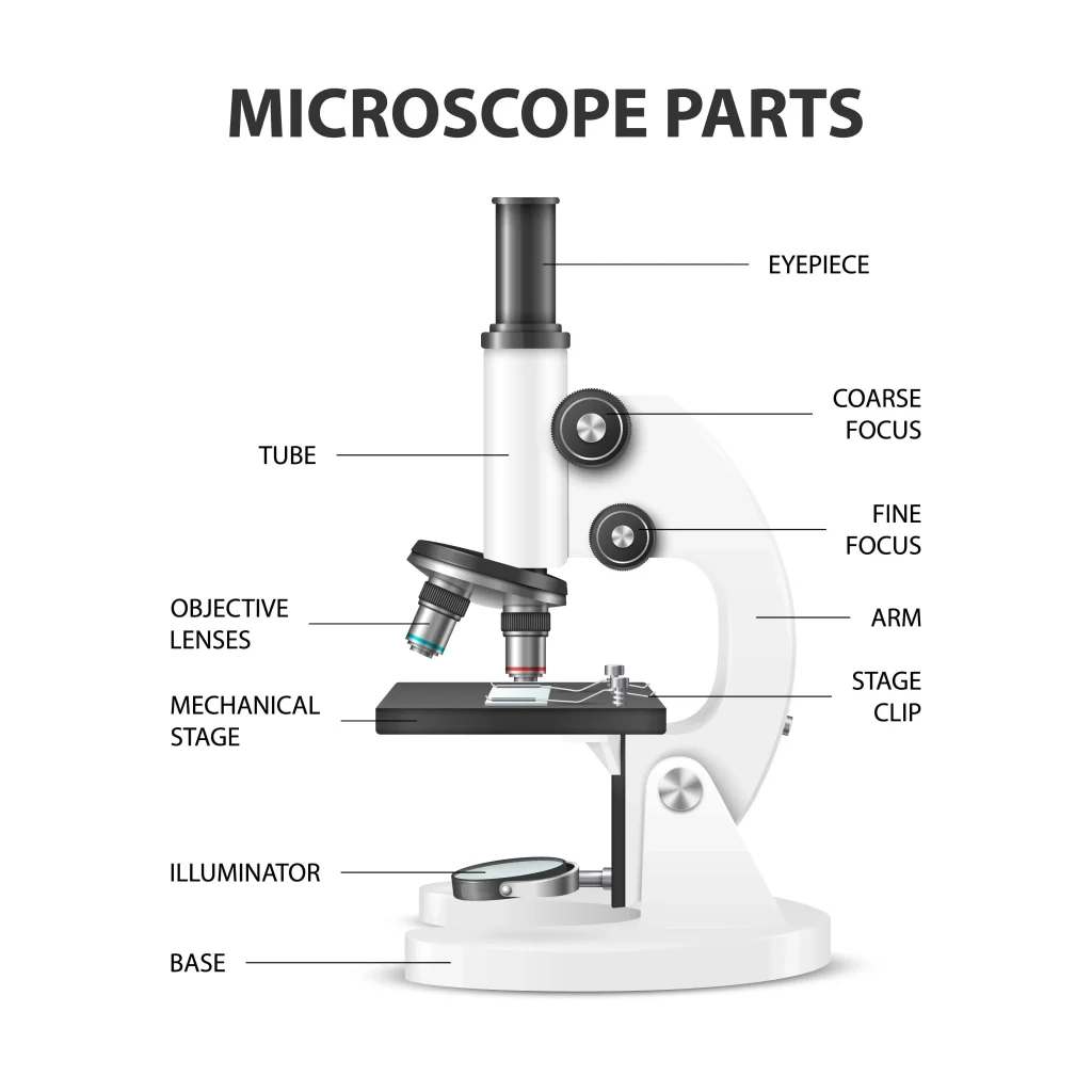
Ocular Lens (Eyepiece)
The ocular lens, also known as the eyepiece, is the upper optical component you look through to observe the magnified image. The ocular lens typically magnifies the image from the objective lens by a factor of 10 to provide the final view.
Microscopes frequently use two ocular lenses with various field of view widths.
Objective Lens
The objective lens is the lower optical element that is closest to the sample. Typically, objectives ranging from 4X to 100X magnification are attached to a rotating turret for simple magnification adjustments.
Together with the ocular lens, objective magnification determines total visual magnification.
Focus Knob
The focus knob raises and lowers the stage to provide a sharp image. While fine adjustments permit precise focusing, coarse adjustment knobs allow for substantial focus variations. The optimal way to view images is with a wide range of depth of field.
Stage
The stage is the surface on which sample dishes or microscope slides are mounted for observation. Using stages enables X and Y positioning to view diverse sample areas.
Stage clamps safeguard samples. Motorized or mechanical stages permit precisely programmed movement.
Stage Clips
Typically, spring-loaded stage clamps are used to secure microscopy samples to the stage surface in order to prevent slides or dishes from shifting during observation. Gentle but constant pressure stabilizes delicate samples without compromising their integrity.
Diaphragm and Condenser
The condenser and diaphragm control the intensity and focus of light particles passing through the sample. The condenser gathers and intensifies the light from the source. The diaphragm regulates the aperture, impacting resolution and contrast.
Illuminator
Below the stage, the internal or external illuminator serves as the light source for transmission and reflection of specimens. Presently, light-emitting diodes (LEDs) are widely used. Adjustable intensity controls the lamp’s brightness.
Arm/Frame
The arm or frame of a microscope connects and positions the optical elements and provides structural support. This primary chassis includes focus gears, lens tubes, wiring routes, and mounting points for stability.
Base
The sturdy base supports the column or arm of the microscope and contains the power supply and illumination controls. Heavy bases prevent instruments from tipping and keep them firmly planted during use.
Functions of Microscope Parts
Beyond physical descriptions, the significant functions that the main parts of a microscope provide are as follows:
Lenses
- Objective and ocular lenses magnify specimens to reveal microscopic details and generate a viewable image. Different lens powers allow for scalability across numerous size ranges.
Focusing Elements
- Coarse and fine focus knobs allow the stage to be moved with precise control to achieve specimen clarity from close to far away. enables the sharpest views.
Sample Stage
- Holds specimens steady for viewing. Allows navigation of the surface of a slide or dish. Maintains sample stability during image capture.
Lighting and Adjustments
- Light from internal or external illumination passes through the sample. Condensers concentrate light. The diaphragm regulates volume and contrast.
Structural Components
- The frame, the column, and the base provide structural integrity and component connections. Secure the microscope for stability while in use.
Types of Microscopes
Various technologies of microscopes are suited for specific applications:
Light Microscopes
- Use visible light and glass lenses to magnify up to one thousand times. View color and detail in living and fixed samples that are transparent. Widely employed in the field of biology.
Electron Microscopes
- Utilize an electron beam and electromagnetic lenses to generate magnifications greater than 10 million X. Reveal cell organelles and proteins as ultrastructures.
Specialized Microscopes
- Developed for specific imaging requirements. Adaptable microscopes include stereo zoom, digital fluorescence, confocal, two-photon, and atomic force microscopes.
Microscopy Methods
Unique microscope technologies enable different imaging techniques:
Brightfield and Darkfield Microscopy
- Brightfield transmits white light through stained or unstained specimens in the presence of a bright background.
- Darkfield scatters light from the periphery to illuminate bright objects against a dark backdrop. Frequently used together to collect comprehensive sample data.
Phase Contrast and Fluorescence Microscopy
- Phase contrast transforms minute differences in sample density into perceptible variations in contrast, as opposed to brightness.
- Fluorescence employs fluorophore tags excited by particular wavelengths to emit colored light, highlighting antibody-labeled cell components.
Confocal and Super Resolution Microscopy
- Through point illumination and pinhole filtration, confocal microscopes eliminate out-of-focus light in order to capture crisp 3D sections.
- Super resolution methods overcome the diffraction limit through techniques like stimulated emission depletion (STED) microscopy.
Microscopy Applications
Microscopes support discoveries across diverse scientific domains:
Biological Imaging
From cells to microbiomes, microscopes enable the visualization of microorganisms, biostructures, and dynamic processes that contribute to the comprehension of life. Essential for pathology, neuroscience, and developmental biology, amongst other disciplines.
Medical Diagnostics
Clinical microscopes enable accurate diagnoses by identifying pathogens, parasites, abnormal cells, and other health indicators. Microscopic examination is used for routine analysis.
Material Science
Microscopy is used to characterize a material’s microstructure, crystallinity, composition, and defects. Guides the development of enhanced-property industrial alloys, ceramics, polymers, and nanomaterials.
Semiconductor Research
Microscopy with high resolution of nanoscale structures such as quantum dots, wires, and transistors enables the design of semiconductors. Critical to the advancement of microelectronics.
Future of Microscope Technology
Ongoing microscope advances expand imaging capabilities:
Novel Imaging Techniques
Emerging techniques such as Bessel beam arrays, expansion microscopy, and AI-enhanced imaging provide new sample views to reveal previously hidden characteristics.
Increased Magnification Power
Next-generation microscopes will shatter the diffraction limit, utilizing novel techniques to achieve atomic resolution nondestructively and image single molecules in real time.
Automation and Quantitative Analysis
Automation of the microscope, correlative workflows, and computational image analysis will facilitate the generation of large amounts of data to quantify phenomena in complex biological systems and diverse materials.
Parts of the Microscope Worksheet
FAQs
What are the parts and functions othe microscopef microscope?
Here is a brief overview of the main parts and functions of a microscope:
Ocular lens (eyepiece) – Magnifies the image from the objective lens and brings it into focus for your eye.
Objective lenses – Magnify and focus the light from the specimen. Different objective lenses provide different levels of magnification.
Stage – Supports the slide with the specimen on it. Can be moved up and down to focus.
Light source – Provides light that passes through the specimen. Often an LED or halogen bulb.
Condenser lens – Focuses the light on to the specimen.
Diaphragm – Controls the amount of light that hits the specimen.
Coarse and fine focus knobs – Move the stage up and down to bring the specimen into focus.
The basic function is to magnify a small object by passing light through it and using lenses to enlarge the image for detailed observation. The ocular lens further magnifies the image.
Who is the father of microscope?
Antoni van Leeuwenhoek
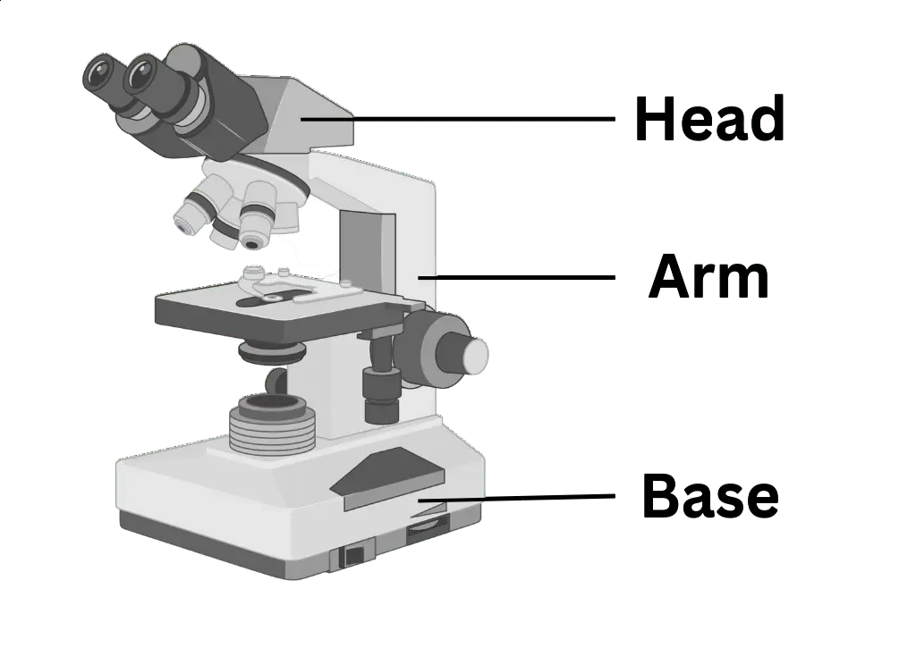
How many parts does microscope have?
The compound microscope has three main structural parts – the head, base, and arm. The arm is the part that connects the base to the head. It supports the microscope head and allows it to be positioned properly for viewing specimens.
The arm can also be used to carry or transport the entire microscope. The base provides stability and a platform on which the microscope rests.
The head contains the optical components like eyepiece, objectives, condenser and light source which work together to magnify the specimen.
So in summary, the arm, base, and head are the three core structural pieces that make up the compound microscope.
What is the function of eyepiece?
The eyepiece, also known as the ocular lens, magnifies the image produced by the microscope’s objective so that it can be viewed by the human eye. This article examines the various types of eyepieces, their components, how they function, and how to use them.
References and Sources
- https://rsscience.com/compound-microscope-parts-labeled/
- Microbiology by Lansing M. Prescott (5th Edition)
- https://www.pobschools.org/cms/lib/NY01001456/Centricity/Domain/349/TheMicroscope-howtouse.pdf
- https://sciencing.com/parts-microscope-uses-7431114.html
- https://www.amscope.com/microscope-parts-and-functions/
- https://cpb-us-e1.wpmucdn.com/cobblearning.net/dist/3/4204/files/2018/08/Parts-of-the-Microscope-103b21p.pdf
- https://biologynotesweb.com/parts-of-a-microscope/